Innovative Technology Expands Applications of Monoclonal AntibodiesーRapid conversion of monoclonal antibodies to multiplexed super-resolution imaging probesー
- Professor WATANABE, Naoki
- Assistant Professor MIYAMOTO, Akitoshi
- Single-Molecule Cell Biology
Overview
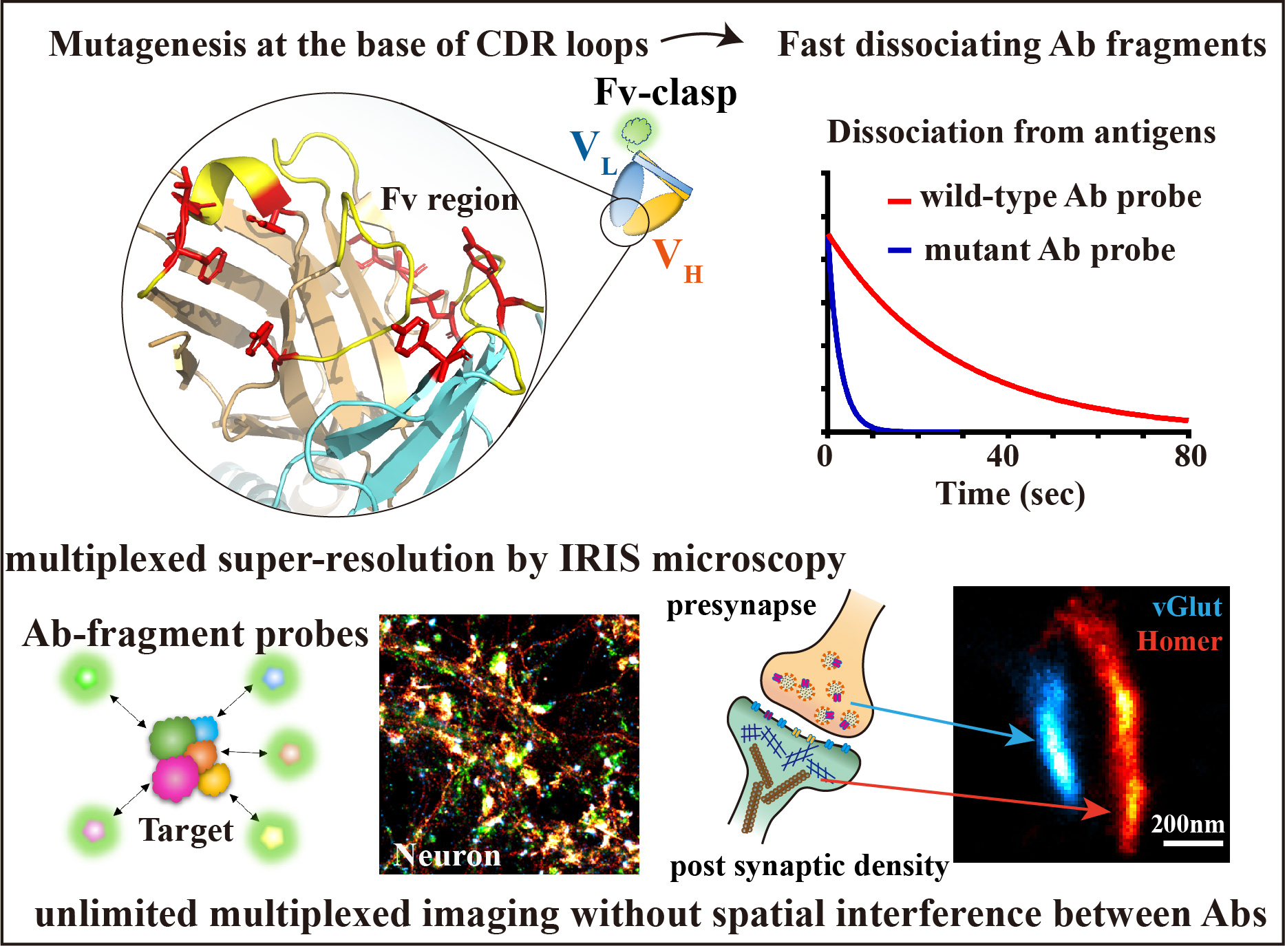
Super-resolution microscopy called IRIS, which uses fluorescent probes that rapidly bind and dissociate to the target molecules, produces high-density, high-fidelity images of molecular distributions that cannot be achieved with conventional antibody staining. IRIS overcomes the “super-resolution dilemma” problem, known for grainy, discontinuous images due to the limited labeling density by conventional antibody staining (Nature Methods 12: 743-746, 2015). However, it was not an easy task for researchers to create IRIS probes for individual targets.
A research group led by Professor Naoki Watanabe of Kyoto University Graduate School of Biostudies (also Professor at Graduate School of Medicine), Assistant Professor Akitoshi Miyamoto, and PhD student Qianli Zhang, in collaboration with Professor Junichi Takagi of Institute for Protein Research, Osaka University, has developed a method to rapidly produce fluorescent probes optimized for the unlimited multiplexed super-resolution microscopy IRIS by modifying existing antibodies. Many proteins are present in thousands to several million per cell. IRIS has the potential to visualize almost all positions of each protein and compare the localization between many types of proteins in a single super-resolved image. It is expected to be a major step toward the practical application and widespread use of IRIS microscopy.
By using the technology developed in this study, a large number of monoclonal antibodies developed for medical and research use can be efficiently converted into fluorescent probes useful for IRIS super-resolution microscopy and other types of multi-antigen detection devices.
This work was published online in Cell Reports Methods on September 20, 2022 (local time).
Researcher’s comment
By utilizing many monoclonal antibodies, that are the assets of mankind, we will be able to greatly increase the variety of molecules that can be visualized with the multiplexed super-resolution microscopy IRIS. Although IRIS requires long image acquisition time, I hope that the “deep field” in the cell will reveal appearance of many functioning proteins entangled with each other. (Naoki Watanabe)
Publication
-
Title
Engineered fast-dissociating antibody fragments for multiplexed super-resolution microscopy
-
Authors
Qianli Zhang, Akitoshi Miyamoto, Shin Watanabe, Takao Arimori, Masanori Sakai, Madoka Tomisaki, Tai Kiuchi, Junichi Takagi and Naoki Watanabe
-
Journal
Cell Reports Methods
Resercher’s information
| Researcher |
Professor WATANABE, NaokiSee faculty information
Assistant Professor MIYAMOTO, AkitoshiSee faculty information
|
|---|---|
| Laboratory | Laboratory of Single-Molecule Cell Biology |
| Laboratory website | http://www.pharm2.med.kyoto-u.ac.jp/2_index.html |